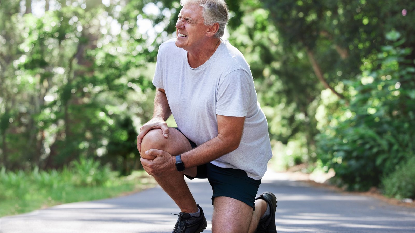
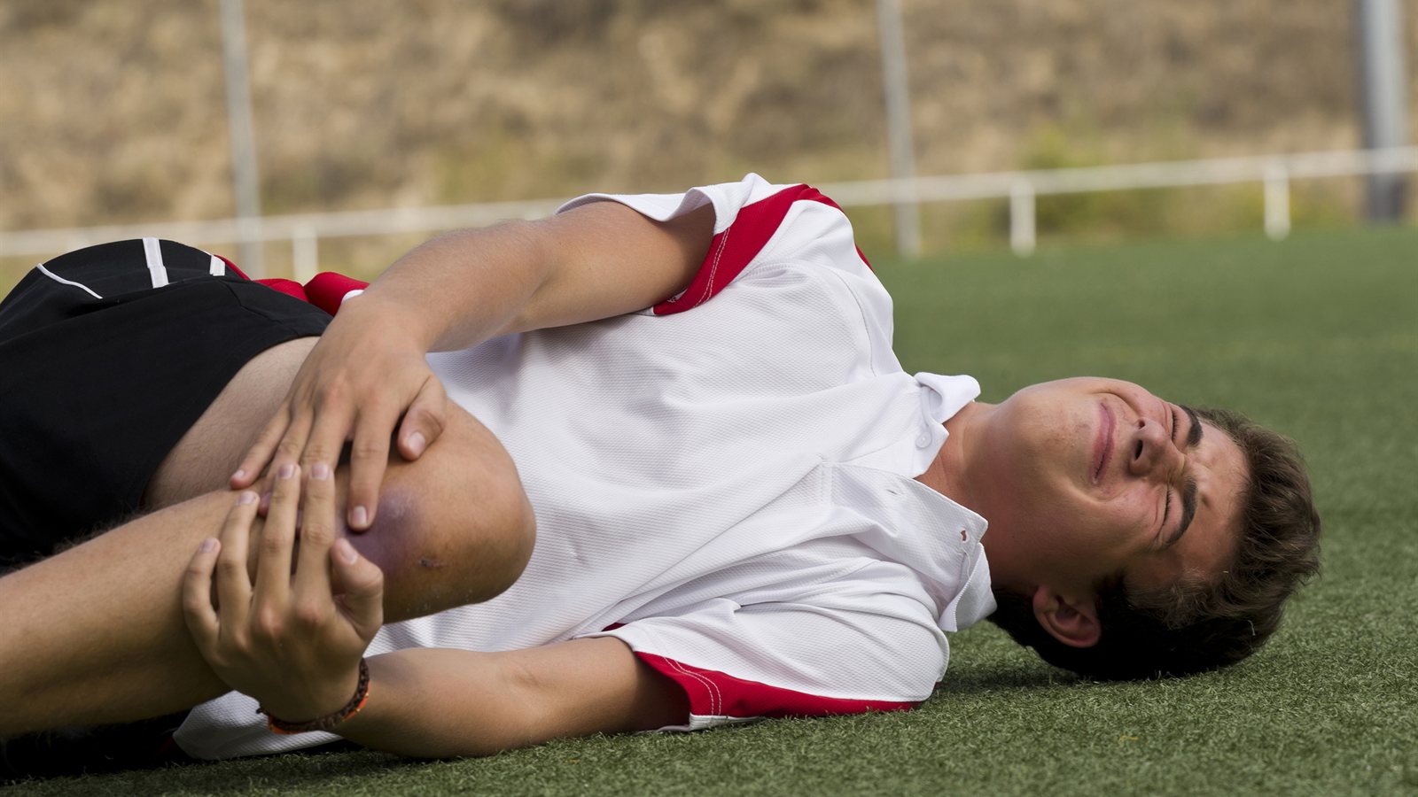
A teenager pivots during a netball game and her knee buckles beneath her. A weekend footballer lands from a jump and feels a pop in his knee. These moments are familiar to families and athletes across Australia, and they often mark the sign of an injury to the anterior cruciate ligament, or ACL.
The ACL is one of the most frequently injured structures in the knee. It plays a central role in joint stability, and when it tears, the effects extend beyond the immediate pain and swelling. Timely, accurate ACL tear treatment by an orthopaedic specialist matters because the decisions made in the first weeks after injury influence long-term joint health, including the risk of future cartilage damage and post-traumatic arthritis.
This guide outlines what an ACL injury involves, how it is recognised and diagnosed, the treatment pathways available, and what to expect during ACL tear recovery. AVA Orthopaedics provides specialist orthopaedic assessment for patients across Australia, with a focus on structured, individualised care. Specialist Advice. Early Direction in what matters to you.
What Is a Knee Ligament Injury and Why Does the ACL Matter?
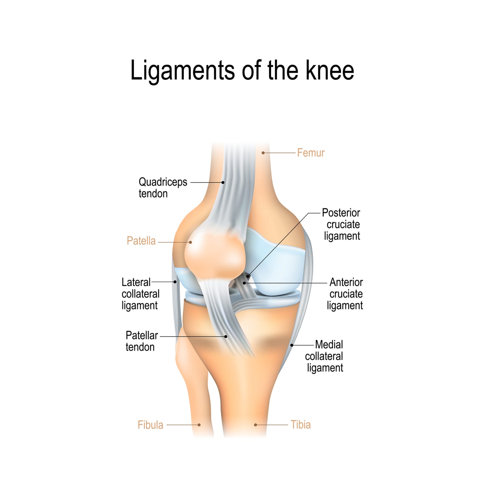
The knee is stabilised by four major ligaments: the anterior cruciate ligament (ACL), the posterior cruciate ligament (PCL), the medial collateral ligament (MCL) and the lateral collateral ligament (LCL). Each contributes to a different aspect of joint control. The two collateral ligaments resist side-to-side forces, while the cruciate ligaments sit within the joint and control forward, backward and rotational movement.
The ACL prevents the tibia (shin bone) from sliding forward on the femur (thigh bone) and provides rotational stability, particularly during pivoting and cutting movements. This is why the ligament is especially vulnerable in sports that involve rapid change of direction, deceleration and jump landings.
A knee ligament injury involving the ACL is clinically distinct from injuries to the MCL, LCL or PCL, though combined injuries are common. The "unhappy triad," for example, involves the ACL, MCL and medial meniscus. Identifying associated ligament, meniscal or cartilage damage is essential to formulating an accurate treatment plan, which is why a thorough specialist assessment is recommended after any significant knee injury.
Who Is at Risk of an ACL Sports Injury?
.jpg)
ACL injuries can occur at any age, but certain groups are at a higher risk. Adolescent female athletes have a notably increased incidence than their male counterparts in the same sports, particularly in netball, soccer and basketball. Adult male contact sport players are also frequently affected, and recreational sportspeople returning to activity after a period away from training are another well-recognised group.
In Australia, the highest rates of ACL sports injury are seen in netball, Australian Rules football, soccer, basketball and snow sports.
Intrinsic risk factors include limb alignment, the shape and width of the intercondylar notch, hormonal influences in female athletes, and a history of previous knee injury. Extrinsic factors include playing surface, footwear, fatigue late in a game, and inadequate warm-up.
An ACL sports injury warrants prompt review by an orthopaedic specialist rather than a watch-and-wait approach. Early assessment by an orthopaedic surgeon (knee/ACL specialist) clarifies the diagnosis, identifies associated injuries and shapes the management pathway from the outset.
Recognising the Symptoms of an ACL Tear
Many patients describe an audible or sensory "pop" at the moment of injury, often accompanied by an immediate sense that something significant has happened inside the joint. Swelling typically develops rapidly within the first few hours, reflecting bleeding within the joint (haemarthrosis) from the torn ligament.
Common symptoms include:
- Sudden pain at the time of injury, often centred deep within the knee
- Rapid swelling within two to four hours
- A sensation of the knee "giving way" or feeling unstable when weight-bearing
- Difficulty fully straightening or bending the knee
- Reluctance or inability to bear full weight
These symptoms can overlap with other acute knee injuries, including meniscal tears, patellar dislocations and tibial plateau fractures. This is why clinical examination and appropriate imaging are needed to confirm the diagnosis.
Emergency department review is appropriate when there is severe deformity, inability to bear any weight, suspected fracture or significant uncontrolled pain. In most other cases, an urgent specialist appointment within the first one to two weeks is the more useful pathway.
How an ACL Tear Is Diagnosed
Diagnosis begins with a structured clinical history. The mechanism of injury, the timing and pattern of swelling, any previous knee problems, current symptoms and the patient's functional goals all inform the assessment.
During a telehealth consultation, the specialist team takes a detailed history and reviews the injury event in depth, including how the knee behaved at the moment of injury, the speed of swelling, and any episodes of instability or giving way in the days that followed. Where patients have undergone clinical examination through their GP, emergency department or physiotherapist, those findings are reviewed alongside the history.
Imaging plays a central role in diagnosis and is particularly well-suited to remote specialist review. Plain X-rays are used to rule out associated fractures, including bony avulsion injuries where the ligament has pulled away with a fragment of bone, and growth plate injuries in younger patients. MRI is the most accurate imaging investigation for confirming an ACL rupture, grading the injury and identifying concurrent pathology. It is particularly valuable for assessing the menisci, cartilage surfaces, bone bruising patterns and the posterolateral corner of the knee.
Where imaging has already been completed, the specialist team reviews the studies directly and discusses the findings with the patient. Where further imaging is indicated, this is arranged as part of the consultation pathway. Hands-on clinical examination, where required, is coordinated through referral to an appropriate local provider.
Engaging an ACL specialist early supports accurate interpretation of imaging, clarifies the diagnosis when multiple structures are involved, and shapes the treatment roadmap from the outset.
ACL Tear Treatment Options: A Personalised Approach
ACL tear treatment is not a single pathway. The most appropriate approach depends on the patient's age, activity level, the degree of instability, associated injuries within the knee, and individual goals for return to work, sport or daily activity.
Broadly, treatment falls into two categories: conservative rehabilitation and surgical reconstruction. The decision between the two is made collaboratively between the patient and the specialist team, taking into account the clinical findings and the patient's circumstances.
Conservative Management: Rehabilitation Without Surgery
Conservative management may be appropriate for lower-demand patients, older adults whose activities do not involve pivoting, and those with partial ACL tears who retain good functional stability.
The goals of non-operative care are to restore quadriceps and hamstring strength, neuromuscular control and proprioception (the body's sense of joint position). A structured physiotherapy programme progresses through phases, beginning with swelling and pain management and advancing to functional retraining.
Bracing may be considered during certain activities, and activity modification is often part of the early stages. Patients managed conservatively are monitored for recurrent episodes of instability. If the knee continues to give way despite rehabilitation, surgical reconstruction may then be considered.
Surgical ACL Reconstruction: What Patients Need to Know
Surgical reconstruction is generally considered for patients with recurrent instability, those with associated meniscal injuries warranting repair, young, active patients, and individuals whose work or sporting demands require a stable knee.
Graft options include autografts (tissue from the patient's own body, most commonly the hamstring tendon or the bone-patellar tendon-bone graft) and, less commonly in younger patients, allografts from a donor source. Graft selection is individualised based on patient anatomy, activity profile and surgeon assessment.
ACL reconstruction is typically performed arthroscopically, through small incisions, under general anaesthesia. Most patients are discharged the same day or following a short hospital stay.
As with any surgical procedure, ACL reconstruction carries risks that are discussed in detail before surgery. These include infection, deep vein thrombosis, stiffness, graft failure, ongoing pain and the possibility of re-rupture. Benefits include restoration of rotational stability and the opportunity to address associated injuries within the joint.
Pre-operative physiotherapy, sometimes called "prehabilitation," is an important part of the pathway. Reducing swelling, restoring range of motion and rebuilding quadriceps strength before surgery is associated with a smoother recovery.
ACL Tear Recovery: What to Expect at Each Stage
ACL tear recovery is a graduated, milestone-based process. Timelines are useful as a general guide, but progression depends on individual response and the achievement of specific functional benchmarks rather than the calendar alone.
Phase 1: Acute Management (Weeks 1 to 2). The focus is on controlling pain and swelling, beginning gentle range-of-motion exercises and activating the quadriceps muscle. Weight-bearing status and the use of crutches are guided by the specialist team.
Phase 2: Rebuilding Strength and Stability (Weeks 3 to 12). Progressive resistance training targets the quadriceps, hamstrings, gluteal muscles and calf. Balance and proprioception retraining are introduced. The psychological aspects of this phase, including frustration with restrictions and the work required to maintain motivation, are recognised as part of the recovery experience.
Phase 3: Functional and Sport-Specific Rehabilitation (Months 3 to 6). Running, cutting, jumping and landing mechanics are reintroduced in a structured way. Sport-specific drills and agility work follow. Objective testing, including limb symmetry index and single-leg hop tests, helps assess readiness. Psychological readiness, including managing the fear of re-injury, is increasingly recognised as an important consideration.
Phase 4: Return to Sport and Long-Term Joint Health (Months 6 to 12 and Beyond). Return to sport is criteria-based rather than time-based, meaning the focus is on demonstrated function rather than the number of months since injury. Ongoing strength maintenance and injury prevention work continues well beyond return to play. The evidence shows a measurable risk of re-injury and contralateral ACL tear, which is one reason continued specialist monitoring and structured maintenance are valued. There is also recognised long-term risk of post-traumatic osteoarthritis, which informs the importance of joint care over the years that follow.
The Role of an ACL Specialist in Your Recovery Journey
An ACL specialist brings focused expertise in the assessment of ACL injuries. This includes detailed knowledge of the pros and cons of conservative vs surgical intervention, and associated injury patterns.
Effective ACL care is multidisciplinary. The team typically includes an orthopaedic surgeon, a physiotherapist with experience in post-ACL injury rehabilitation, and at times, a sports psychologist to support readiness and confidence.
Ongoing specialist review at key milestones, rather than only at the beginning and end of recovery, allows treatment to be adjusted as the patient progresses. Telehealth and online consultations have expanded access to specialist orthopaedic input for patients in regional and remote areas of Australia, as well as for those balancing recovery with work and family commitments.